
The Micro-Incision Revolution: How Minimally Invasive Orthopedic Surgery Redefined Recovery
Traditional open-bone surgeries with massive scars and months of bed rest are becoming things of the past. Driven by rapid technological shifts in robotic assistance, high-definition visualization, and specialized miniature tools, minimally invasive orthopedic surgery allows surgeons to fix joints, repair ligaments, and stabilize fractures through incisions no larger than a keyhole. This educational guide breaks down how the technology works, why it drastically slashes recovery timelines, and what candidates can expect.

For decades, the prospect of undergoing orthopedic surgery evoked images of substantial hospital stays, long structural scars, and grueling, months-long recovery periods. The traditional "open" approach required large incisions to allow surgeons a direct line of sight and hands-on access to bones and joints. However, a profound paradigm shift has arrived. Today, orthopedic medicine focuses heavily on muscle-sparing and tissue-preserving methods. Through micro-incisions, advanced fiber-optic cameras, and ultra-precise robotic arms, surgeons can now perform complex joint reconstructions and spinal repairs with minimal disruption to the surrounding healthy tissue. For patients, this means less pain, significantly lower infection rates, and a remarkably fast return to daily life.
The Micro-Incision Revolution: How Minimally Invasive Orthopedic Surgery Redefined Recovery
For decades, the prospect of undergoing orthopedic surgery evoked images of substantial hospital stays, prominent structural scars, and grueling, months-long recovery periods. The traditional "open" approach required large incisions to allow surgeons a direct line of sight and hands-on access to bones and joints.
However, a profound paradigm shift has arrived. Today, modern orthopedic medicine focuses heavily on muscle-sparing and tissue-preserving methods. Through micro-incisions, advanced fiber-optic cameras, and ultra-precise robotic arms, surgeons can now perform complex joint reconstructions and spinal repairs with minimal disruption to the surrounding healthy tissue. For patients, this means less pain, significantly lower infection rates, and a remarkably fast return to daily life.
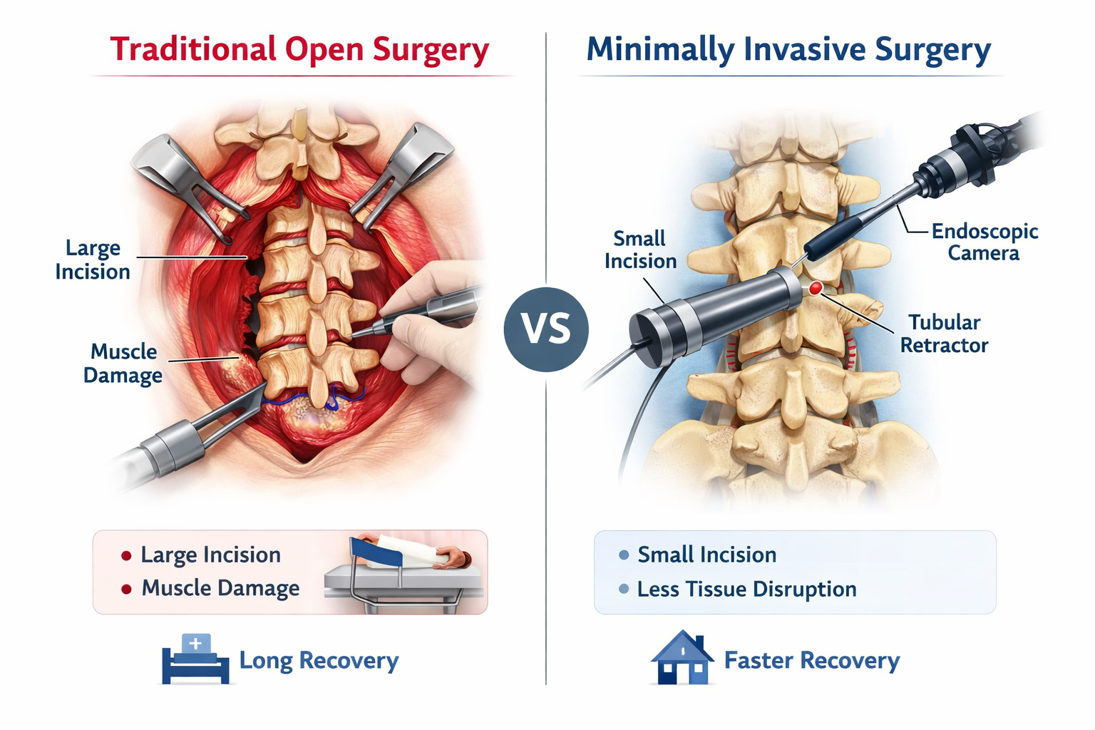
The Mechanics: How Keyhole Surgery Works
The magic behind modern tissue-preserving procedures lies in two core technologies: arthroscopy (using a miniature camera inside a joint) and specialized, long-shafted micro-instruments.
Instead of cutting through large muscle groups to expose a damaged knee or shoulder, the surgeon makes a few tiny punctures. One puncture accommodates a high-definition camera called an arthroscope, which projects a magnified, 3D view of the surgical field onto high-resolution monitors. The remaining punctures serve as entry ports for specialized instruments engineered to cut, shave, or anchor tissues with sub-millimeter precision.
Precision Enhanced: The Rise of Robotics
- Surgical precision has entered a new era through the integration of robotic-assisted joint replacement systems.
- Pre-Operative 3D Modeling: Before making a single cut, advanced software transforms standard CT scans into a personalized 3D roadmap of your unique bone structure.
- Intraoperative Guidance: During surgery, robotic arms provide real-time feedback, preventing the surgical tool from deviating even a fraction of a millimeter outside the pre-planned safety boundaries.
- Systems like the Mako technology pictured above do not replace the surgeon; rather, they act as a high-tech shield. The robotic arm ensures that cutting tools only interact with the precise areas targeted for implant alignment, leaving healthy bone and tissue completely untouched.
The Recovery Advantage: Why Less Trauma Means Faster Healing
Dr. Kshiteez Puri
Orthopedic SurgeonOrthopedic Surgeon in Nepal specializing in joint replacement, sports injury treatments, and advanced fracture care.